Search here!

Custom Search

About Me

Wednesday, November 18, 2009

The art of letting go

“We often fool ourselves and say that it’s love

only cause when it’s gone, we end up being lonely

So how are we to know that it just wasn’t so

That we have to let each other go…”

Its over… she’s gone…

Why do we have to part while the love is still there? Why do we have to suffer? Why do we have to cry when someone bids goodbye? Why do beginning has an end? Why do we have to meet only to loose in the end?

There are questions left unanswered, words left unsaid, letters left undone, songs left unsung, love left unexpressed, promises left unfulfilled.

In a relationship, one of the hardest things to do is saying goodbye and letting go. It is as hard as breaking crystal because you’ll never know when you’ll be able to pick up the pieces again. More often than not, they who go feel not the pain of parting: it is they who stay behind that suffer because they are left with memories of a love that was meant to be… a love that was.

“There are times when we share precious moments

but later realized they are only stolen moments

So how are we to know that it’s just wasn’t so

That we just let each other go…”

At the beginning and at the end of a relationship, we are embarrassed to find ourselves alone. Unfair as if they may seem, but that’s the way love goes. That’s drama, the bitter sweet and risk of falling in love. After all, nothing is constant but change, everything will eventually come to an end without asking when, without knowing how without why. And we must forget not because we want to but because we hate to.

In letting go, sorrows come not as a single spy but in battalion. It seems that everywhere you go, every song you sing, every turn of your head, every move of your body, every beat of your heart, every blink of your eyes and every breath you take, it always reminds you of her. Its like a stab of knife, torture in the night. Funny how the whole world becomes depopulated when only one person is missing. Just imagine there are four billion people on earth and yet it seems you only feel lonely and empty without the other.

I don’t know if it’s worth calling an art, but letting go entails special skills sparkle with considerable space and time. Time heals all wounds but it takes a little push on our part. Acceptance plays a part. Not all wishes do come true. No all love stories end with “and they live happily ever after.”

Sometimes we have to part because of circumstances beyond our control. We have to suffer if it would mean happiness for others. We have to cry temporarily to let go of the pains. Every beginning has its end like every dawn has its dusk. It’s something we can’t control, something we have to live up.

Its over… she’s gone…

But life has to go on. Goodbye doesn’t mean forever. There will always be a place and time where questions will be answered, words will be spoken, letters will read, poems will be recited in the night, songs will be sang in harmony, love will be expressed in solitude and promises will be fulfilled. Somewhere. Somehow. Someday.

“if loving you is all that means to me,

and being happy is all I hope you’ll be.

If loving you must mean,

I really have to set you free…”

- spongebob

Thursday, September 10, 2009

Plant and Animal Dissection Handbook

Plant and Animal Dissection Handbook

by ROLLY B. CAIDIC
University of the Philippines Open University
Diploma in Science Teaching
Major in Biology


Ready? Get Set… Dissect!
DSCN4356
What do you know about plants and animals? I’m sure you have an idea about its parts and how it works but can you tell exactly where, why and how those parts function? In this handbook, you are going to learn biology by dissecting a euphorbia and a dog; you will need to work and think like a biologist. Most important of all, you will want to ask yourself the questions that a biologist asks:
What is this part?
How does it function?
Why?
These are the questions that most scientist ask when they experiment. When a scientist experiment, he tries to discover a new fact or idea. Sometimes he may try to prove something people already know or think they know.
You will find dissection activities here that can be done by using ordinary euphorbia plant or our askal breed dogs and tools that are easily found. Just remember, a scientist watches his work carefully and thinks about what he is doing. He isn’t afraid to try something new or do it in a different way.
The experiments here are not fully described, so do them as you think they should be done.
DSCN4374
The pictures will be helpful in giving you starting ideas. Don’t be discouraged if you fail occasionally. That’s the way to experiment and discover. So are you ready? Get set… GO Dissect!


PLANT SPECIALIZATION

Crown of Thorns
Local Name in Filipino: Euphorbia
Scientific Name: Euphorbia milii var. splendens (uu-for' bee-ah mill' ee-ii splen' dens)
5586
Kingdom Plantae – Plants
Subkingdom Tracheobionta – Vascular plants
Superdivision Spermatophyta – Seed plants
Division Magnoliophyta – Flowering plants
Class Magnoliopsida – Dicotyledons
Subclass Rosidae
Order Euphorbiales
Family Euphorbiaceae – Spurge family
Genus Euphorbia L. – spurge
Species Euphorbia milii Des Moul. – christplant
Variety Euphorbia milii Des Moul. var. splendens (Bojer) Ursch & Leandri

INTRODUCTION

The joy of planting Euphorbia
What’s this craze over euphorbia? Why are people going gaga over this thorny plant? Look around your town and you will notice that many people, whether rich or poor have euphorbia plants displayed in their front yards. Some are planted in clay pots, black plastic pots and some are planted in lowly tin cans. It’s always a pretty sight to behold - euphorbia plants teeming with pretty flowers.
It’s a staple in local gardens. Aren’t they come in different colors of yellow, pink, orange, white, red in varying shades, some are combination of colors? It’s also available in different sizes, from cute button-like blossoms to inch-sized flowers. You can also spot them being sold by ambulant vendors during market day. It seems that this craze has been going on for some time now and vendors are making good money from it.

Why Euphorbia?
As the picture shows, you can observe a lot of things from euphorbia. I think that euphorbia easily caught the fancy of plant lovers because it blooms abundantly all year round. I heard someone say that it’s not greedy with its flowers. How true! Just look at the plant when it’s in bloom. It has so many flowers, that sometimes there are more flowers than the leaves. And yes, the flowers in different sizes, variety of colors, and unique formation of petals are so captivating. You can also admire the flowers longer coz they don’t easily wilt. Another reason I suppose is that it’s very easy to propagate and grow.

Plant it yourself!
DSCN4376
The first collection of euphorbia plants you can have is from your friends’ and neighbors’. They could give you some stems cut from the “mother” plants and plant them in pots. But wait! The newly planted cutting shouldn’t be watered for at least a week they call this technique in tagalog as “binibihag”. Why do you think they do this? Another helpful advice is that it should be exposed to sun; so it will yield an abundant bloom. This is a low maintenance plant as it has high tolerance for heat. Do we need to water it more often? What are the other planting practices should you consider in planting this kind of succulent deciduous shrub?

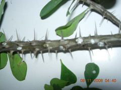
MAJOR EXTERNAL STRUCTURES
DSCN4379
What are the major parts of a plant? What are their functions?
Watch out for the thorns! Ask an adult to help you get a plant sample of euphorbia. Use hand gloves or any protective gear when handling the plant.
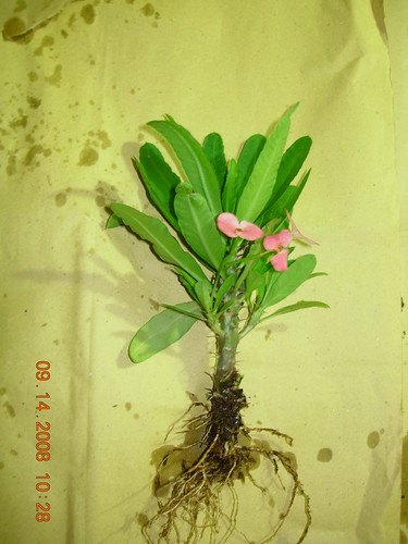
Observe the picture showing the major organs of a plant. Can you see these on your euphorbia plant? What are the differences between the parts shown on the drawing and the parts of your plant sample? You can use a ruler or a tape measure in measuring your specimen. How can you describe each part of euphorbia plant?


STEM DISSECTION
What’s inside the succulent stems of euphorbia plant?
DSCN4362 copy
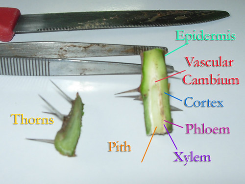
Remove all the leaves and thorns of your plant sample. Carefully cut the widest stem part crosswise. Use longer forceps and knife and hand gloves when doing this. Do you see a white – milky sap coming out of the stem? What is this sap for? Those can be very dangerous and toxic so avoid getting it on your skin and especially on your eyes! Can you see sap like this coming out from the other parts of the plant?
Wipe off the sap with a piece of cloth or paper. Then slice the stem lengthwise. How do you describe the internal parts of the stem? Can you see different layers inside the stem? What layers of the stem are saps coming from?
Identify those parts of the stem by comparing it with the diagram below.


FLOWER DISSECTION
DSCN4383
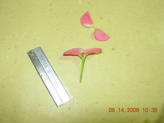
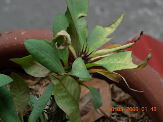
Did you know that the brightly colored ‘petals’ we see on euphorbia are actually modified leaves called ‘bracts’? The flowers which are called cyathia (singular: cyathium) are in the center, quite small and uninteresting looking. What do you think is the flower’s function? Why do you suppose the bracts of flowers of euphorbia are so colorful, uniquely shaped? Do they have some special purpose for that?
By using a scalpel or an ordinary blade cut the flower of the euphorbia plant lengthwise. Be careful not to damage the central part, remember that’s the actual flower and important parts are there. Can you identify the parts of the flower by comparing it with the picture below?


MORPHOLOGY OF EUPHORBIA
DSCN4359 copy
PARTS OF THE STEM

DSCN4366
Stems are structures which support buds and leaves and serve as conduits for carrying water, minerals, and sugars. The three major internal parts of a stem are the xylem, phloem, and cambium. The xylem and phloem are the major components of a plant’s vascular system.
The vascular system transports food, water, and minerals and offers support for the plant. Xylem vessels conduct water and minerals, while phloem tubes conduct food. The vascular systems of monocots and dicots differ. While both contain xylem and phloem, they are arranged differently. In the stem of a dicot such as the euphorbia, the vascular system forms rings inside the stem. The ring of phloem is near the epidermis or external cover of the stem and is a component of the epidermis in mature stems. The xylem forms the inner ring; it is the vascular cambium in herbaceous plants.
The cambium is a meristem, which is a site of cell division and active growth. It is located between the xylem and phloem inside the bark of a stem and is the tissue responsible for a stem’s increase in girth, as it produces both the xylem and phloem tissues.

PARTS OF THE FLOWER
The sole function of the flower, which is generally the showiest part of the plant, is sexual reproduction. Its attractiveness and fragrance have not evolved to please man but to ensure the continuance of the plant species. Fragrance and color are devices to attract pollinators that play an important role in the reproductive process.
The pistil is the female part of the plant. It is generally shaped like a bowling pin and located in the center of the flower. It consists of the stigma, style, and ovary. The stigma is located at the top, and is connected to the ovary by the style. The ovary contains the eggs which reside in the ovules. After the egg is fertilized the ovule develops into a seed.
The stamen is the male reproductive organ. It consists of a pollen sac (anther) and a long supporting filament. This filament holds the anther in position so the pollen it contains may be disbursed by wind or carried to the stigma by insects, birds or bats.
Sepals are small green, leaflike structures on the base of the flower which protect the flower bud. The sepals collectively are called the calyx.

CYATHIUM
Cyathium_cross1_ies
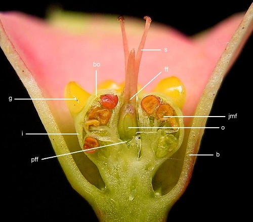
The very special pseudanthia in the genus Euphorbia (Euphorbiaceae) are known as Cyathia. A cyathium consists of:
• five (rarely four) bracteoles. These are small, united bracts, which form a cup-like involucre. Their upper tips are free and in the beginning cover the opening of the involucre (like the shutter of a camera). With these alternate:
• five (1 to 10) nectar glands, which are sometimes fused.
• one extremely reduced female flower standing in the centre at the base of the involucre, consisting of an ovary on a short stem with pistil, and surrounded by:
• five groups (one group at the base of each bracteole) of extremely reduced male flowers, which each consist of a single anther on a stem.
The flower-like characteristics of the cyathia are underlined by brightly coloured nectar glands and often by petal-like appendages to the nectar glands, or brightly coloured, petal-like bracts positioned under the cyathia.

END NOTE
Congratulations! You are now an experienced biologist. If you have kept notes on your work you will be able to check each idea that you worked on. You can even design and write about your own experiments too. Look around! Try dissecting other parts of the plant or animal you’re working on. Keep trying and you can discover a lot more.
This handbook is designed to delight and challenge the young biologists. This will help him apply methods of scientific research to find answers to these questions for himself. All the dissections can be conducted with a plant or animal and other indigenous materials.

ANIMAL SPECIALIZATION
Filipino Dog
Local Name in Filipino: “Aso”/ “Askal” (Asong kalye) / “Aspin” (Asong Pinoy)
practice dissect copy
Kingdom Animalia (animals)
Eumetazoa (metazoans)
Bilateria (bilaterally symmetrical animals)
Deuterostomia (deuterostomes)
Phylum Chordata (chordates)
Craniata (craniates)
Subphylum Vertebrata (vertebrates)
Superclass Gnathostomata (jawed vertebrates)
Euteleostomi (bony vertebrates)
Class Sarcopterygii (lobe-finned fishes and
terrestrial vertebrates)
Tetrapoda (tetrapods)
Amniota (amniotes)
Synapsida (synapsids)
Class Mammalia (mammals)
Subclass Theria (Therian mammals)
Infraclass Eutheria (placental mammals)
Order Carnivora (carnivores)
Suborder Caniformia (caniform carnivores)
Family Canidae (coyotes, dogs, foxes, jackals,
and wolves)
Genus Canis (dogs, jackals, and wolves)
Species Canis lupus (gray wolf)
Subspecies Canis lupus familiaris (dog)

INTRODUCTION

The Love of Filipinos for Dogs
cutie
Do Filipinos share a special bond with Dogs? The dog is one of the most popular animals in the world. It was among the first animals to be domesticated, or trained for use by humans. It is characterized by loyalty, friendship, protectiveness, and affection. Therefore, dogs are also known as man's best friend.
The bond between dogs and people has existed for thousands of years, and dogs are still an indispensable part of the lives of farmers, ranchers, hunters, police and customs officers, and many people with disabilities, among others. In the Philippines dogs serves as “bantay” or guard of the house. They are treated just like one of the members of the family.
An askal is a mixed-breed (mongrel) dog in the Philippines. The name is a Tagalog-derived contraction of asong kalye (street dog). It is applied to mongrels due to their often stray nature. These dogs are often more resilient than pure-bred ones.
Unlike other dog breeds whose origins can be readily traced, the askal’s roots are unclear. But we do know that they have been around for a very long time. Documents dating back to the late 17th century show evidence of the existence of these dogs in Philippine soil. A photograph taken in 1904 even shows an Igorot tribesman with his hunting dog. Whether these dogs are native to the Philippines or introduced to the country by foreign traders and explorers is still debatable.

Physical Characteristics
Domestic dogs vary widely in appearance, particularly in size. The Shih Tzu, for example, is 20 to 28 cm (8 to 11 in) in length and weighs 4 to 7 kg (9 to 15 lb). The Irish wolfhound is at the other end of the scale, measuring about 71 to 94 cm (about 28 to 37 in) at the shoulder and weighing up to about 61 kg (about 135 lb). Coat color, length, texture, and pattern also vary greatly. The muzzle may appear shortened, as in the Pekingese, or elongated, as in the Doberman pinscher. Limbs are relatively short in the basset hound and dachshund, but long in the greyhound. Ear shape and carriage also vary, but these characteristics may be influenced by a dog owner’s decision to crop, or cut, the ears to make them stand up. Some dogs, notably the chow chow, even have a naturally blue-black tongue.
The askal is suitably built for the tropical climate of the Philippines. Its short and practically non-shedding coat helps the dog withstand the hot, harsh summers in our country. These dogs are, quite contrary to belief, intelligent. Their street-smart ways can attest to that. Leave another breed of dog to fend for itself on the streets and I can assure you, it cannot adapt as well as an askal can. It is also fallible to say that native dogs cannot be trained. An askal can hold its own against any breed. It is not only foreign breeds that can be taught to perform tricks. With patience and the proper training techniques, any breed of dog has the potential to learn, and an askal is no exception.


Owning Your Own Askal
Every Pinoy probably grew up with dogs around them. But the plight of askals is pitiful. Thousands live off the streets because their owners either got tired of taking care of them or because the dogs have become too much of a burden. Owning a dog entails a lot of responsibility. The decision to adopt a dog should be made carefully because it is a serious commitment that can last for several years. Small dogs may live 12 or more years, although very large dogs typically have a shorter lifespan, sometimes as brief as 8 years. Before owning a dog, potential owners should examine their lifestyle, living accommodations, and plans for the dog. Other decisions should include who, in the case of a family, will care for the dog and whether the family or individual owner will have enough time, attention, and money to meet the dog’s needs.
Askals, like any other dogs, need food, water, and most importantly, a good home. The streets are not good homes for them. They could easily get infected with canine viruses such as parvo, distemper and rabies in the streets. The high incidence of human fatalities due to rabies in our country should not be blamed on askals, but on human negligence. And sadly, hundreds of homeless askals are sent to dog pounds every year where they are put to sleep.

DISSECTION
Anatomical Direction
Before beginning a dissection, it is important to have an understanding of some of the basic directional terminology associated with the dissection of specimens. Some of these terms include proximal, which means toward the body, and distal, which means to move away from the body. Other important anatomical directions are indicated below.

Key Anatomical Directions
433034801_f3a7fe913d

Dissection Safety Rules
Proper safety procedures when working with dissection tools and specimens is of greatest importance. Some safety rules to engage in when dissecting specimens are as follows.
• Follow all instructions given by your teacher.
• Inform your teacher of any illness as a result of exposure to chemicals used in specimen preparation.
• Avoid contact with preservative chemicals. Rinse the specimens completely before dissection.
• Know where the eye-wash fountain is if needed.
• Wear safety goggles to prevent the splashing of any chemicals into the eyes.
• Properly mount dissection specimens to dissecting pan. Do not dissect a specimen while holding it.
• Handle scalpel or razor blade (safety edged) with extreme care.
• Always cut away from your body and away from others.
• Never ingest specimen parts.
• Never remove specimens or specimen parts from the classroom -- until the dissection is completed all parts of the dissection must remain within the dissecting pan.
• Properly dispose of dissected materials.
• Store specimens in as directed by your teacher.
• Clean up the work area and return all equipment to the proper place when the dissection is completed.
• Wash hands after each dissection.
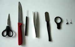
Dissection Equipment
The pictured dissection equipment from left to right is (1.) a teasing or dissection needle which used to pull apart muscle tissue, (2) dissecting scissors which are used to cut through tissue, and (3) a scalpel, which is a knife used to slice through and cut tissue.

ANIMAL DISSECTION
The dissection of animals is important for many reasons. It helps in the learning about the internal structures of animals. It also allows students to learn how organs and tissues are interrelated. Another purpose of dissection is to allow the comparison of organisms in terms of their organs and relative complexities. While many good simulations of dissections may be observed, it seldom can replace the benefits of the actual participation in an actual dissection.
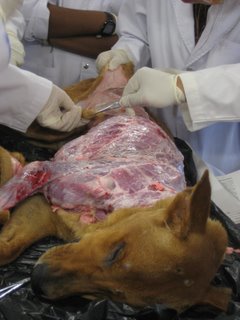
Usually the dissection procedure involves tying the organism down firmly on the dissection pan, cutting the organism open on its ventral side (as pictured below), and pinning its tissues and muscles back to observe its internal organs. Different teachers may have their own preferences in terms of their emphasis on the tissues and organs to be observed in a dissection.
Dissection_018

Skinning

How can you describe the skin? What is its function? Lay the cat on its back on a flat, easy to clean surface. Taking a small pair of sharp scissors, start to make a cut at the lowest point of the front of the ribcage, in the centre. Do you feel the natural hollow where your bottom ribs join the sternum? That is where you need to start cutting. Snip a shallow cut. Insert the rounded blade of the scissors just under the surface and cut up the way, in a straight line to the neck. From the neck, cut in a line towards the right corner of the mouth. Repeat the process by cutting down along the belly to reach the pubic bone (part of the pelvis). The long cut you have just made is called the ventral cut. Snip down the inside of each leg to the knee and then down to the dew claw in a smooth, clean line. At the dew claw, make a cut around the circumference of the leg, taking care not to nick any tendons.
Reaching into the ventral cut, carefully tease the skin away from the underlying muscle or bone until the skin has been freed from the body of the dog, but is still held at the head and anus. You will notice a cotton-like, fibrous material that ties the skin to the flesh. This is called the superficial fascia. Be careful to only remove the skin and not the tissue. Particular problem areas will be the flanks, the neck, the shins and particularly the head, when you get to it. Take your time and ease the skin away in the difficult areas.
Now that the skin is mostly free, it's time to start cutting again. Using the scissors, start to cut down from the pubic bone past either side of the sex organs. Cut around either side of the perineum (genitalia and anus) towards the tail, thus effectively circling this part of the dog’s anatomy. Then cut the skin between the tail and the anus, thus leaving a ring of fur around the perineum. Make a cut down the length of the tail and peel the skin away.


Muscular System
Hind Limb (medial)
Carefully remove the skin by pulling it down and off the end of the lower leg. You can cut it off if it is too difficult to remove, but do not cut any muscle fibre. The yellowish-white material under the skin is fat. Is there more fat in the thigh or the leg?

Now that the skin and fat are off, you can see the muscle – the ‘meat’. Examine the muscle and separate the bundles of muscles with your fingers. What is the function of muscles? Begin the dissection by inserting your thumb into the muscle of the lower leg. You will need to push quite hard through the shiny lining (called fascia) that is over the muscle, but it will give way at the natural separations between the muscle bundles. Continue separating the muscle into bundles by forcing your thumb and fingers through the muscle until you are able to see several separate bundles. At both ends of the muscles, you will see the strong, white cords, called tendons. What are tendons for? Can you differentiate it from muscles and other organs in the hind limb?

Thoracic Cavity
907968722_c640234ac7
The thoracic cavity is separated from the abdominal cavity, which contains the gastrointestinal tract, by the thoracic diaphragm. The thoracic cavity contains the great vessels, the heart and the lungs, and some smaller structures.
To expose the contents of the thoracic cavity, first use a scalpel and made a transverse incision anterior to the lowest point of the front of the sternum in order to avoid cutting into the abdominal cavity. The incision was caudal to the ribs that attached to the sternum and ran from the right side of the ventral thorax to the left side of the ventral thorax. Cut across the area posterior to the clavicles and remove the cut ribcage. Can you identify the lungs and diaphragm? What organ system do they belong? The dog has right and left lungs, each covered in parietal and visceral pleura. There are how many lobes in the lungs? Can you identify them and their locations?
Carefully remove the left lung, to be able to see the dorsal aorta, esophagus, vagus nerve, and azygous vein. What organ system these organs belong to? What are its functions? The most medial of the four is the descending aorta. Lateral (left side) to it is the left vagus nerve, which is located superior to the esophagus. The esophagus is situated deep to the vagus and medial to the azygous.

2365731230_c28bfe52e5
Digestive System

The dissection part is very simple. Make an incision on the ventral side of the dog starting at the diaphragm and continuing all the way down to the base of the tail, opening up the abdominal cavity. The organ that is most cranially situated in the abdominal cavity is the liver. What is the important function of live? What are the secretions produced by liver?
The stomach is situated caudal to the rib cage in the left part of the abdominal cavity. The stomach has a few different roles in the digestion process. What are those functions? Can you identify the stomach and liver from the rest of the organs?
The structure that takes up most of the space in the abdominal cavity is the intestine. How can you describe the intestines? The intestine is usually divided into the small intestine and the large intestine. Can you identify the two types? What is its importance in digestion of food?
Excretory System
Before the excretory system can be examined, the gut tube, and all of its associated organs (liver, spleen, gall bladder, pancreas, etc.) must be removed. Remove all of the internal organs together being careful not to damage the urogenital organs and all of their associated vasculature and ducts. The region dorsal to the guts should be cleaned.
The kidneys are “retroperitoneal” or embedded in the abdominal wall and it is surrounded by a relatively thick pad of fat. Remove the tissue and the fat surrounding the kidneys. Be careful not to damage any of the vessels running to and from the kidneys, the ureters running from the kidneys to the bladder, or the adrenal glands, which sit on the cranial end of each kidney. Can you describe the kidneys? What are its major function?
Follow the ureters caudally to the bladder. The bladder is ventral to the uterus and rectum in the pelvis. If the two ureters carry urine produced in their respective kidneys where is it stored? How does the bladder works upon urination? What part does the urine leaves the body?

Ovaries and Uterus

Female dogs, like all female mammals, have two small ovaries. They are located caudal to the kidneys and craniolaterally to the uterus. Each has its own vascular supply and is attached to the body wall via the ovarian ligament. The ovaries are closely associated with the uterus (and indeed appear attached to it) and lie at the apex of each of the two uterine horns.
The uterus of the dog is very different than that of the human (in fact, the cat’s uterus is closer to the typical mammalian condition) in that it has two distinct horns. In the pregnant cat, each of these horns can contain multiple developing fetuses with zonary placentas. Each fetus has its own placenta, chorion and amnion and its own pouch-like region of the uterus.
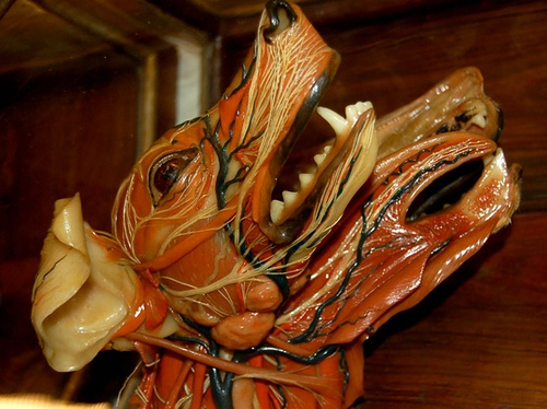
Brain

Begin this dissection by scraping away the temporalis muscle from the anterior portion of the skull. Once the skull was visible, we used scissors to cut around the perimeter of the skull, making sure not to damage the brain. It is difficult to cut through the skull because the bone was harder and thicker than most of the other bones in the dog’s body. What is the function of the skull?
Remove the top of the skull and notice the dura mater coming off attached to the inside of the skull. What do you think is this for? Using a probe, gently lifted the brain out of the head. Then cut the brain stem, fully freeing the brain from the head. Inside of the brain case, see the optic chiasm where the optic nerves cross and then enter the brain. Still attached to the brain was one of the two optic nerves. What organ is the other end of optic nerves and what is its function?
To divide the brain into its right and left hemispheres, use a scalpel cut down the longitudinal fissure of the brain. The interior view of the brain revealed the third and fourth ventricles as well as the corpus callosum. In addition, a distinct separation between the pons and medulla oblongata are visible. Can you identify these parts of the brain? What do you think are their functions?

LAST WORD
Why do we dissect animals and plants?
We dissect to provide a way for us to learn about the internal and external anatomical structures of animals and plants. By taking a hands-on learning approach, we can get a real sense of the relationships between organism’s structures. Drawings and pictures are not enough. The true feeling of being in the field and actual dissection is very different than by just imagining it.
For many students however, dissections can be very disturbing. Online dissections provide the means to experience actual dissections without all of the mess. But the real deal is not there. It is my idea not to provide all the answers in this handbook. Questions are provided to suggest things to watch for and ways to look for them to precede them with independent investigations.

REFERENCES
Bailey, Regina (2008). Virtual Cat Dissection. About.com.: Biology. Retrieved September 14, 2008, from http://clk.about.com/?zi=1/XJ&sdn=biology&cdn=education&tm=569&gps=
59_803_1276_577&f=00&su=p897.2.336.ip_&tt=2&bt=0&bts=1&zu=
http%3A//bio.bd.psu.edu/cat/
Buckley, James M. Jr. (2003). Dissection. Oswego City School District Regents Exam Prep Center. Retrieved September 10, 2008 from www.regentsprep.org/Regents/biology/units/laboratory/dissection.cfm
Dazee (2006) Euphoria over Euphorbia. Dazee Writes About. Retrieved September 12, 2008 from http://dazeee.i.ph/blogs/dazeee/2006/04/26/euphoria-over-euphorbia/
Faucon, Philippe (2005) Crown of Thorns. Desert Tropicals. Retrieved August 7, 2008, from http://www.desert-tropicals.com/Plants/Euphorbiaceae/Euphorbia_milii.html
Jacqueline (2007) Splendid Euphorbia milii in exotic and bizarre colors! John & Jacq’s Garden. Retrieved September 13, 2008, from http://www.jaycjayc.com/euphorbia-milii-crownofthorns/
Mitchell, Bob (2005) Euphorbia milii var. splendens. St. Andrew’s Botanic Garden. Retrieved August 7, 2008, from http://www.st-andrews.ac.uk/~gdk/stabotanic/nov05pom.htm
Pignatti, Sandro (1982) Flora d'Italia, Edagricole, Bologna. ISBN 8850624492. Retrieved September 14, 2008 from "http://en.wikipedia.org/wiki/Cyathium"
Tan, Michael (2007). "Askal". Philippine Daily Inquirer. Retrieved September 14, 2008, from http://opinion.inquirer.net/inquireropinion/columns/view_article.php?article_id=62750
University of Arizona (1998) Botany: Plant Parts and Functions. Arizona Master Gardener Manual. Retrieved Sept. 14, 2008 from http://ag.arizona.edu/pubs/garden/mg/botany/plantparts.html
University of Michigan (2008) Canis lupus familiaris dog. Animal Diversity Web. Retrieved Sept. 14, 2008 from animaldiversity.ummz.umich.edu/site/accounts/information/Canis_lupus_familiaris.html
Vincentz, Frank (2006) Image:Cyathium cross1 ies.jpg. Wikimedia Commons. Retrieved August 7, 2008, from http://commons.wikimedia.org/wiki/Image:Cyathium_cross1_ies.jpg
Weil, Anne (2002). Comparative Mammalian Anatomy. Duke University Biological Anthropology & Anatomy. Retrieved September 14, 2008, from www.baa.duke.edu/companat/BAA_289L_2004/index.htm

Wednesday, August 5, 2009

INTERNET ACTIVITY NO. 21


CELLULAR RESPIRATION

Welcome aboard everyone!
We will have our very first ONLINE ACTIVITY here. I will require you to have some basic knowledge about computers and internet. At any rate, you may seek coaching from your classmates or just tap someone’s shoulder besides you and don’t hesitate and don’t be scared to ask. = )

Answer all the Guide Questions at the end part of this activity. Then before signing out here please give me your feedbacks. How was your experience in using internet? Does this activity helped you? Did you experienced any problems> What are those? I need those answers so I may troubleshoot some inadequacies in using this type of technology. There is a big big room for improvements here. I am always looking forward for your constructive criticisms, suggestions or comments.

You have two options in passing your Internet Activity:
1. Send your ANSWERS to the Activity Sheets online through my email address: rollycaidic@yahoo.com
Indicate your Complete Name and the Activity No. as your subject… or
2. Print your answers on short bond paper then pass it to me personally on Tuesday.

Enjoy learning everyone!


INTERNET ACTIVITY NO. 21
CELLULAR RESPIRATION

Converting Food to Usable Energy
Cellular respiration is a complex series of chemical reactions that harvests the energy trapped in the bonds of glucose sugar molecules and converts it into a chemical form that can be stored for later use. Humans and other animals obtain glucose sugar molecules from food, such as the fruits and grains shown here, while plants create glucose sugars in the process of photosynthesis. Essential to the survival of most organisms on Earth, cellular respiration yields the energy that makes our bodies function properly.


Procedure:
I. Click on the following websites. Then answer the following Guide Questions.

here are some videos i got from youtube.com

II. Guide Questions:
A. In what form is energy stored in humans?
B. Why do organisms need to store energy?
C. Write the equation for aerobic cellular respiration
D. What are the stages of aerobic respiration?
E. Describe the following:
a. Glycolysis
b. Kreb’s Cycle
c. Electron Transport System
F. Differentiate anaerobic cellular respiration from aerobic respiration
G. What is the site of aerobic cellular respiration in all eukaryotic cells?

Friday, June 19, 2009

more planting trials


My first few attempts were in vain though. The plants died after three months much to my disappointment. How ignorant I was back then! I didn’t even asks my co-teachers how many times a week should I water it and where should it be placed. Few weeks have passed and this is now what my plant looks.


This is now my second attempt to propagate a euphorbia plant. Just in time for my plant dissection handbook, I will be able to use it as well during my dissections. Now I have a lot of questions in mind to ask the seller of euphorbia plants in our weekly “tiange”.


1. What is the breed of this euphorbia plant? Its imported she said…
2. Every when should I water it? Every week she answered…
3. Do I need to apply fertilizers on it? Yes, monthly would do. Do not overdo it…
4. Where should I place my plant? Near well lighted places.
5. Can I took it out of the soil then bring it back after my examination? Yes but do not water it right away. Wait for 2 days after then water it.
6. Why? So that the plant can adopt to a new soil and it would not drowned.
7. How much is it? Only 25 pesos. Different varieties are more expensive because of the petals.
8. Can I propagate it? Yes, one tip from me, choose the plants which has plenty of stems. You can cut these off later and plant, thus increasing your collection.


Isn’t it great? Now I’m caught in the euphorbia fever too!

Thursday, June 18, 2009

the euphorbia fever

Few months ago, I noticed some of my cp-teachers which are plant lovers are requiring drop-to-be students to bring euphorbia plants to our school as their project. Yeah it’s a laughing stuff at school normally students wouldn’t buy teachers’ wants. They thought it was a joke bringing plants to save their failing grades but my co-teachers were not joking! They really badly wanted to propagate this plant to their gardens. As soon as I am aware of this, that’s the time I noticed most of the front yards are starting to change their old plants to euphorbia. Until I realized euphorbia paint the town of Lumban red and pink.

So for my plant specialization, I chose this because of its abundance to my locality. I bought my first plant last March 2008. Brightly and bloody colored red flowers and heaps of green leaves there is. Then school days start and I forgot to take good care of the plants. As soon as this come to my attention, flowers begins to fall one at a time. I thought the plant were just adjusting to its new environment.

One time I got the chance to look at it closer. As I am getting close I noticed leaves were also diminishing! Mostly were already decaying on the soil and some had jagged margins. I tried to look underneath the leaves and to my surprise two caterpillars are devouring my plants! I hurriedly pick a stick and took it out of my sight. Well too late, leaves are badly damaged and stems were all left out. I pull out the complete fertilizers I bought together with the euphorbia then I observe it after a few days later. These are some of the pictures I took for my documentation.

this photo was taken after i took my euphorbia plant away from the garder in front of our yard because i thought caterpillars that ate my plant were from the other plants. i placed my euphorbia plant in front of my window next to where i could always observe it. i also apply fertilizers to it and water it every after two days.

Monday, June 8, 2009

About my Euphorbia Plant

I bought my euphorbia plant last February 2008 for the flower pots in front of our house. I chose this kind of plant because summer days are coming and I have learned from my co-teachers that these relatives of cactus grows best in sunny and dry places. Another thing that I consider in buying euphorbia is its low maintenance characteristics. It doesn’t require full attention and even watering is not on regular basis. Another is because it is always in full bloom most of the time.
Upon choosing the euphorbia I pick the ones with bright red color-flowers. It’s like red roses from a far. The flowers are very attractive and the plants look healthy. The sales man said it is a hybrid. I also bought 2 packs of complete fertilizers. The gardener said it would also help improve the flowering of my plant.
here is a short background information about my plant specialization:


Crown of Thorns


Scientific Name: Euphorbia milii var. splendens Des Moul.

Synonym: Euphorbia splendens

Kingdom Plantae – Plants
Subkingdom Tracheobionta – Vascular plants
Superdivision Spermatophyta – Seed plants
Division Magnoliophyta – Flowering plants
Class Magnoliopsida – Dicotyledons
Subclass Rosidae
Order Euphorbiales
Family Euphorbiaceae – Spurge family
Genus Euphorbia L. – spurge
Species Euphorbia milii Des Moul. – christplant
Variety Euphorbia milii Des Moul. var. splendens


The family Euphorbiaceae is large with about 8,100 species found throughout the world, excepting the Arctic region. Euphorbia itself is a very large genus with over 2000 species. They are characterised by having a milky latex which can cause burns and indeed some may be carcinogenic. The xerophytic species most often have spines and our plant this month - Euphorbia milii var splendens - is no exception. Surprisingly, since it does not need too much watering and, especially the fact that it flowers most of the year, but particularly in the winter months, it has become a cherished house plant for sunny dry situations.

Euphorbia milii var splendens, commonly called Crown of Thorns, is a straggly, spiny shrub bearing bright red bracts and, given a chance, will grow to 2m. Euphorbia milii was named by Des Moulins in 1826 and, while not a succulent, can withstand long dry periods when the plants will shed their leaves. The stems are furrowed and furnished with stout, tapering spines about 2cm long. The leaves occur at the top of the shoots and the branched inflorescences grow from the leaf axils. The variety splendens is simply a larger form than the species itself and occurs naturally. In fact there is considerable variation in the wild in south-west Madagascar and several have been named, including a yellow flowered form, also grown for the pot plant industry.

Of course, the related Poinsettia - Euphorbia pulcherrima - from Mexico, is a major player in the Christmas pot plant industry. These will be on sale from the Friends in December.

Many of the African species of Euphorbia have developed in parallel evolution with the Cactaceae of the Americas. This can be seen in the two borders of the Xerophytic (Succulent) House. (The information board explains the details.) Euphorbia milii var splendens has been pot grown at the Garden for many decades and was planted in the African border when the glasshouse was redesigned about 20 years ago.

Cultivation.
Euphorbia milii var splendens demands a bright, sunny situation and, while it can withstand temperatures as low as 45'F (7.5'C), it is best not to let the temperature fall below 50'F (10'C). A well drained compost is essential, for the roots will not tolerate soggy conditions. In fact the plants are better kept drier in the winter time.

Propagation.
Summer cuttings of the current years growth taken about May or June will root in heat. When taking the cuttings it is best to dip the cut ends in charcoal to stop the latex flow before inserting into a sandy compost.

Position.
Euphorbia milii var splendens is growing in the Xerophytic House on the right at the entrance from the Corridor.

References:

Mitchell Bob, Euphorbia milii var splendens, Retrieved August 7, 2008, from http://www.st-andrews.ac.uk/~gdk/stabotanic/nov05pom.htm

Crown of Thorns, retrieved August 7, 2008 from http://www.desert-tropicals.com/Plants/Euphorbiaceae/Euphorbia_milii.html

Monday, March 16, 2009

my cranial outputz, his cardiac throbz


Sadness fills my empty space as i glare through his heart's summons.
Longing to ease his agony somehow.
I drenched with him in midnight rain. Perilous love puts everything in jeopardy.
I crave to witness the sun shines back at him.
Exorbitant friendship is all I could offer.
Hoping this would tranquil his wounded heart...
For my buddy, whose heart is at apathy of sheer love.


***
Never loose your grip with love for it will find you no matter what. If your heart is nowhere to be found just wait until time let it flies back into you.


***
Monotony of life builds unbalanced being. It entomb one's self in lampoon of hapiness. Friends cease to ease this shanty of isolation. I thank you for taking this part.


***
Choosing chances are only limited to those who conceived of loosing. Never quit in life for it must go on no matter how hard it is. May you have more sucess in life.


***
As i track my way to school. Tracing those paths that once we've taken. Leaving my nest for i have to strugle for life. Until this afternoon we'll cross our paths once more.


***
Memories are engravings of a heart made of gold. For we are the artist that effluence shinning smiles. Every rivulet of happy moments spurt out into an ocean of treasures. May this day carved another impressions on gleaming heart.


***
I may not at your side at times of sadness. I may not right there to wipe away those tears but one thing i can assure you... Every heart aches you keep inside crashes me from within, every tears falling down from your face drowns me tremendously, every agony and pain you feel devours me slowly as well.


***
An empty heart is what he is... Strugling to find a relationship that's true. Like an ocean of scapes with an endless path. Compass his heart to break the hiatus of scarce. He's a scavenger of love, life and friendship. Take every chances of it and cherish them all.

Monday, February 2, 2009

Nung maconfine si nanay

Bata p ako nung mamatay si tatay mga grade 3 sa pagkakantanda ko at malaki pagkawala nya sa aming pamilya..

Sa musmos kong pag akap sa aming kalagayan nagkaroon ako ng takot para sa aking nanay.. Tila ginulat ako sa katotohanang maari itong mangyari sa kanya o sa kahit na sinong aking mahal sa buhay..

Ayaw kong aminin sa sarili kong mangyayari ito pwedeng ngayon, pwedeng bukas o pwedeng sa susunod na panahon pero sa aking pagkukubli sa isip ay lumalabas naman sa aking kilos at gawa..

Tuwing gabi sinasadya kong di agad matulog at bagkus ay hihintayin ko munang mauna matulog ang aking nanay.. Sa kanyang pagkakahiga at mahimbing na pagkakatulog ay dahan dahan kong lalapitan at tititigan ang kanyang dibdib..

Tanong ko pa sa sarili ko noon kung humihinga ba siya? Tumataas baba ba ang kanyang dibdib senyales na siya ay maayos n humihinga? Di pa ako makukuntento at dahan dahan akong lalapit at itatapat ang aking darili sa kanyang ilong upang maramdaman ang hangin n lumalabas pasok sa kanyang baga.. Tinangka ko pang pakinggan ang pagtibok ng kanyang puso ngunit bigo akong magawa ito sapagkat natatakot akong magising si nanay.. Di ko alam ang isasagot ko kung sakaling mahuli nya ako sa ginagawa ko..

Kasabay noon ang aking pagtitig ng mariin sa itsura ng aking ina at tila kinukumbinsi kong masigla at buhay n buhay ang aking nanay sa mga sandaling iyon.. Ilang minuto ko ito ginagawa gabi gabi bago matulog noong bata pa ako.. Tila pilit kong kumbinsihin ang sarili kong di n pwede maulit ang nangyari sa aking tatay at sa murang edad ko ay iyon na ang tanging paraan kong magagawa upang masiguradong di na maaring mangyari uli ang katotohanang tumatakot sa akin..

Simula noon namulat n ako sa katotohanang ang buhay ay mayroong hanganan.. Binuksan nito ang aking murang isip sa reyalidad na walang kasiguraduhan ang bukas at ang mga susunod na araw.. Nabatid kong mas mahalaga ang kasalukuyan kumpara s hinaharap at mas dapat kong pahalagahan ang pagkakataong ngayon na aking tinatamasa..

Sa murang isipan kong iyon lahat bumungad sa aking ang mga karanasang pilit magpahinog sa aking katauhan at gayundin kung paano ko tingnan ang buhay..

Sa pagkakataong ito ay nakaharap muli ako sa aking nanay.. Tila bumalik ako sa aking pagkabata at parang musmos n nakatitig sa kanyang pagkakahiga sa kama ng hospital.. Bumabalik ang multo ng aking nakaraan.. Muli kong kinukumbinsi ang sarili kong may magagawa na ako.. Di na ako tulad ng dating napakamurang edad at halos walang magawa kundi ang tumitig at magbantay sa pagkakahimbing na tulog ng aking nanay..

Siguro nga ngayon ay mas matapang na ako at mas siguradong kayang harapin ang hinaharap.. Nagbago na nga ako ngayon pero isa parin ang katotohanang di ko malulusutan.. Ito ay ang reyalidad na hindi nagbabago ang panahon.. Ang aral ng kahapon ay buhay na buhay at nagbabanta sa akin..

Ginigising ako ng aking ulirat na maaring di pa huli ang lahat.. Mayroon pa akong magagawa sa upang iliko ang hamon ng buhay.. Nanlalamig ang aking talampakan at parang magpapawis ang aking kamay.. Kilos! Kabog ng aking dibdib.. Kilos! Sigaw ng aking isip.. Kilos! utos ng aking katawan..Nagkakasundo ang tatlong bulong sa aking kalooban.. Kailangan ko nang kumilos at gumawa ng paraan.. Hindi simpleng pagkakataong lamang ito na mapunta sa hospital ang aking nanay.. Ito ay isang maagang pagbabadya ng maaring kaharapin ko balang araw..

Yayakapin ko na ang hamon ng kasalukuyan... paghahandaan ko na ang banta ng hinaharap.. Marahil totoong walang kasiguraduhan ang bukas ngunit kaya ko pa itong baluktutin at iligaw..


Sent from my iPhone

Tuesday, January 27, 2009

Evolution Hoax?!: Embryonic Development


One day while preparing some PowerPoint slides about embryonic development for my students I stumbled into one of those site where it explains that pictures I was very familiar with based on the books I have were all hoax!? I was surprised and realized I was somewhat misleading my students on the topic of evolution and embryonic development.

Well just like Lamark’s theory of evolution, we couldn’t change the fact that some “hypotheses” exists just to give notion that there may be some good points and also to give us point of comparison between the other theories such as Darwin.

On this matter, generations of biology students including me, may have been misled by a famous set of drawings of embryos published 123 years ago by the German biologist Ernst Haeckel. They show vertebrate embryos of different animals passing through identical stages of development. But the impression they give, that the embryos are exactly alike, is wrong, says Michael Richardson, an embryologist at St. George's Hospital Medical School in London. He hopes once and for all to discredit Haeckel's work, first found to be flawed more than a century ago.

On the brighter side, it’s always to know a thing which is out of the ordinary books. Our reference materials may not be sufficient enough for these but it’s important that we gather information from different sources and not sticking out on things that our eyes could only see.

Here’s the article:

HAECKEL AND THE VERTEBRATE ARCHETYPE

A recent paper by Michael Richardson and his colleagues (1997) has corrected a major misunderstanding among developmental biologists and evolutionary biologists which can be traced back to Ernst Haeckel. Haeckel (1874) had claimed that members of all vertebrate classes pass through an identical evolutionarily conserved "phylotypic" stage. Until this new paper appeared, it was assumed that Haeckel was correct and that there was a particular stage of development that was identical to all vertebrates. Only later in development would specific differences appear. Interestingly, there was some discussion as to what exactly this stage was (Richardson , 1995). This conserved stage was sometimes considered the neurula stage (Wolpert, 1991), the "pharyngula" stage (characterized by the branchial arches; Ballard, 1981), the tailbud stage (Slack et al., 1993), or the stages between those of headfold and tailbud (Duboule, 1994). Figure 1 shows one of Haeckel's representation of vertebrate embryology. The top row shows that all the vertebrate embryos (no matter what path they took to reach this stage) have a stage where they appear almost identical.


FIGURE 1. Haeckel's 1874 version of vertebrate embryonic development. The top row shows an early stage common to all groups, the second row shows a middle stage of development, and the bottom row shows a late stage embryo. Groups from left to right are: fish, salamander, turtle, chicken, pig, cow, rabbit, and human. (Adapted from Gilbert, 1997.)

But Haeckel's drawings are wrong. Photographing actual embryos at these stages, Richardson and colleagues show that Haeckel's drawings are oversimplified to the point of obscuring important differences between classes of vertebrates. The Richardson et al. paper does not dispute that there is a highly conserved embryonic stage among the vertebrate classes. Indeed, at the late tailbud stage, vertebrate embryos of most all classes possess "somites, neural tube, optic anlagen, notochord, and pharyngeal pouches." However, these authors do criticise the notion that this stage is nearly identical in all species and that differences beteen the classes can be resolved only after subsequent development. Rather, they discover significant differences between groups. Size is one distinctive marker. The scorpion fish embryo is 700 microns long at the tailbud stage, while the mudpuppy salamander measures some 9 millimeters. Heterochrony is another problem. In some species of direct developing frogs, and in monotreme mammals, limb buds are already present at the tailbud stage, whereas in other species, these are not seen until significantly later. Birds are characterized by their prominent mesencephalon. Whereas most amniote embryos have a heart by this stage, the zebrafish does not. (Teleosts such as zebrafish are even the exception to the rule mentioned above. They eventually possess a notochord, somites, pharyngeal pouches, etc., but they do not have the pharyngeal pouches until after the tailbud stage). Some of these differences are depicted in Figures 2 and 3.


FIGURE 2. Selected embryos shown at the "phylotypic" stage. (A) Sea lamprey (jawless cyclostome fish, Petroyzon) with nearly identical pharyngeal pouches and heart that is caudal to the pharynx; (B) electric ray (cartilagenous chondrichthes fish, Torpedo) with nearly identical pharyngeal pouches and a pronounced hindbrain/midbrain flexure; (C) sterlet (bony Osteichthyes fish, Acipenser) where the pharyngeal pouches have not yet formed; (D) frog (direct -developing anuran amphibian Eleutherodactylus) which has hindlimb buds at the tailbud stage and only two pairs of aortic arches; (E) pond turtle (chelonian reptile, Emys) where there is no craniocaudal rotation (torsion) to the embryo, whereas the same stage of the chick (avian) embryo (F) shows pronounced torsion; (G) brush-tailed possum (marsupial mammal, Trichosurus) where there is (in contrast to the cartilagenous fishes) large maxillary and mandibular processes. In ther cat (eutherian mammal, Felis) there is a similar size difference between the anterior and posterior pouches, although their appearance differs from that of the marsupials. (After Richardson, 1997.)

FIGURE 3. Photographs of a fish, amphibian, reptile, mammal, and bird embryo at the "phylotypic" stage. Photographs courtesy of M. Richardson.

Interestingly, this knowledge appears to be "old hat" among German biologists. Haeckel's drawings were not trusted (see Goldschmidt, 1956), and Haeckel was accused of scientific fraud by a university court in Jena, where he worked and by other embryologists, as well (see Hamblin, 1997; Richardson et al., 1997b). Yet, the idea that early vertebrate embryos are essentially identical has survived. I think there were two reasons for the survival. First, Haeckels' illustration was reproduced in Romanes' (1901) Darwin and After Darwin. From here, the illustration entered Anglophone biology, "sanitized" from Haeckel. Second, the picture can be used (as it has been in several developmental biology books, including my own [Gilbert, 1997, p. 254]) to illustrate von Baer's principles rather than Haeckel's biogenetic law. K. E. von Baer had noted that the general features of a large group of animals appear earlier in the embryo than do the specialized features. Indeed, von Baer wrote:

"The embryo of the mammal, bird, lizard, and snake and probably also the turtle, are in their early stages so uncommonly similar to one another that one can distinguish them only according to their size. I posess two small embryos in spirits of wine, embryos whose name I neglected to note down, and I am now in no position to determine the classes to which they belong. They could be lizards, small birds, or even very young mammals."

Darwin quoted this (it was Thomas Huxley's translation) although he misattributed the story to Agassiz rather than to von Baer (see Richards, 1992 for an analysis). The notion of development proposed by von Baer is still used as a general approximation of certain developmental phenomena. Certainly, the early embryos of certain vertebrate classes are very similar (for instance, the chick and mouse twenty-somite embryos), and the processes of somitogenesis, limb formation, axis generation, etc. are probably conserved throughout the vertebrate groups).

Similarities and differences

What the Richardson et al. paper tells us is that we have to be attentive to the differences as well as to the similarities. A bird embryo is not the same as a mammalian embryo. We should not assume identity, even at a stage where all vertebrate embryos seem to pass. The Richardson et al. paper can be read as a critique of the recent priority of similarities over differences.

The concept of homology enables one to celebrate the differences or the similarities between two structures. Whether one emphasizes the similarities between our forelimb and a bird's wing or the differences between them depends on what you are describing. Comparative anatomy--with its Aristotelian and Cuverian interest in relating structure to function--usually emphasizes the differences. Morphology--with its Platonic and Geoffroyan interest in the underlying unities of structure--usually focuses on the similarities.

As Joseph Needham noted, embryology has swung between these two poles during different stages of its morphogenesis. In the late 1800s, the morphological tradition prevailed, and the similarities between developmental stages in different organisms constituted some of the best evidence for classification (see Nyhart, 1995; Bowler, 1996). Thus, the discovery of the Nauplius stage of the barnacle showed that it was a modified crustacean, and the notochord-containing tadpole of the tunicate demonstrated its affinities with the chordates. However, after the 1920s, embryology was no longer a major support for evolutionary biology, and the comparative anatomy tradition came to predominate. Until the 1980s, embryology was extremely descriptive. Each organ was seen to develop differently from any other organ, and each species was seen to develop differently from any other species. Indeed, embryology was defined (by one of its practitioners, E. G. Conklin) as a "lawless science" because generalities could not be made from the observations of animal development. When embryology underwent its anagenic transition to become developmental biology, the similarities among organisms were again emphasized. The similarities were now posited on the molecular rather than the morphological or cell lineage level. The 1990s has seen a remarkable celebration of the similarity of molecular processes throughout the animal kingdom. Homologous genes abound (the Hox genes, fringe , tinman, and Pax6 being seen to specify the anterior-posterior axis, the limb, the heart, and the eye, respectively, of organisms as diverse as insects and flies). Even signalling pathways are seen as being homologous both within a developing organism and between organisms. Thus, the neural tube in vertebrates and insects are seen as being formed through the same interactions of the "same" proteins, even though one neural tube is dorsal and the other ventral. The Richardson et al paper reminds us that despite these similarities, differences are also important, especially if one is thinking in terms of the relationships between development and evolution.

The Richardson et al paper is a also plea for comparative anatomy and less reliance on "model systems." This critique of model systems echoes a recent plea from Leo Buss, Rudy Raff, Jessica Bolker, and James Hanken who observe that model systems converge on several factors (see Bolker and Raff, 1997; Hanken, 1993): they all can develop in the laboratory and are thus relatively free of environmental factors effecting their morphogenesis; they are all organisms which segregate their germ line extremely early; they develop rapidly, are small as adults, and they have short generation times.

The Richardson et al. paper does a great service to developmental biology. As they mention, there is renewed interest in evolutionary developmental biology, and in view of the conservation of developmental mechanisms, we need to re-examine the extent of variation in vertebrate embryos. There is more variation than had been assumed, and these variations foreshadow important differences in the adult bodies. This is important because we need to know how conservative the early embryonic stages actually are.

References:

Ballard, W. B. 1981. Morphogenetic movements and fate maps of vertebrates. Amer. Zool. 21: 391 - 399.

Bolker, J. A. and Raff, R. A. 1997. J. N. I. H. Res. 9: 35 - 39.

Bowler, P. J. 1996. Life's Splendid Drama. U. of Chicago Press, Chicago.

Duboule. D. 1994. Temporal colinearity and the phylogenetic progression: a basis for the stability of the vertebrate Bauplan and the evolution of morphologies through heterochrony. Development (suppl.) 1994: 135 - 142.

Gilbert, S. F. 1997. Developmental Biology. Fifth ed. Sinauer Associates, Sunderland, MA.

Gould, S. J. 1977. Ontogeny and Phylogeny. Belknap Press, Cambridge, MA

Haeckel, E. 1874. Anthropogenie oder Entwickelungsgeschichte des Menschen. Engelmann, Leipzig.

Hambin, T. J. 1997. Haeckel's drawings. Times (London), 18 Aug. 1997.

Hanken, J. 1993. Model systems versus outgroups: alternative approaches to the study of head development and evolution. Amer. Zool. 33: 448 - 456.

Nyhart, L K. 1995. Biology takes Form: Animal Morphology and the German Universities 1800-1900. University of Chicago Press, Chicago.

Richards, R. J. 1992.The Meaning of Evolution. U. of Chicago Press, Chicago.

Richardson, M. K. 1995. Heterochrony and the phylotypic period. Dev. Biol. 172: 412 - 421.

Richardson, M. K., Hanken, J., Gooneratne, M. J. Pieau, C., Raynaud, A., Selwood, L., and Wright, G. M. 1997. There is no highly conserved embryonic stage in the vertebrates: implications for current theories of evolution and development. Anat.Embryol. 196: 91 - 106.

Richardson, M. K., Hanken, J., Selwood, L., Wright, G. M., Richards, R. J., Pieau, C., and Raynaud, A. 1997b) Haeckel, embryos, and evolution. Science 280: 983 -984.

Romanes, G. J. 1901. Darwin and After Darwin. Open Court, london.

Slack, J. M. W., Holland, P. W. H., and Graham, C. F. 1993. The zootype and the phylotypic stage. Nature 361: 490 - 492.

Wolpert, L. 1991. The Triumph of the Embryo. Oxford University Press, Oxford.


Your Horroscope